


 720-777-0123
- Your Visit
-
Research & Innovation

It starts with a Q:
For the latest cutting-edge research, innovative collaborations and remarkable discoveries in child health, read stories from across all our areas of study in Q: Advances and Answers in Pediatric Health.


Pediatric Cardiac MRI, CT Scan Tests and 3D Heart Models
We are one of the largest programs in the country treating patients with heart problems from before birth through adulthood, with exceptional outcomes.
Doctors use magnetic resonance imaging (MRI) and computed tomography (CT) scans to diagnose possible heart problems and to monitor children diagnosed with heart conditions. Images from these tests help doctors understand the health of your child’s heart and plan possible interventions and treatments.
What is cardiac MRI
MRI is a type of imaging test that can see parts of the body in detail using magnetic waves. With a cardiac MRI, we can see detailed images of the chambers of the heart and large blood vessels while it’s beating.
The most common type of heart MRI uses a contrast dye provided to the patient through an IV in their arm that makes it easier to see some details of the scan. If a contrast isn’t necessary, then it won’t be used as part of the test.
By using a heart MRI, a doctor can understand how well a patient’s heart is pumping and how blood flows through the heart and vessels.
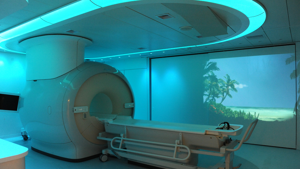
What to expect from a cardiac MRI
Cardiac MRIs usually take between 1 hour and 90 minutes to perform.
During the test, your child will be asked to hold their breath for about 10 to 15 seconds at a time to minimize how much the motion of breathing impacts the pictures of the heart. Children over six years old who can lie still and hold their breath are encouraged to do the cardiac MRI without general anesthesia. However, some children who have difficulty remaining still can receive general anesthesia to help them during the test.
Before your test, we help make your child feel as comfortable as possible with the use of our expert child life specialists, pediatric nurses, technologists and child-friendly MRI rooms. Children can watch a movie during the test to help them relax and pass the time. We have a collection of movies at all our locations, or feel free to bring your child's favorite.
Learn more about what to expect when getting an MRI at Children’s Hospital Colorado.
What is a 4D flow MRI?
4D flow MRI is an innovative MRI sequence that allows clinicians to visualize their patient’s heart and all its neighboring blood vessels in 3D while the heart is beating in real-time. Our multi-departmental heart imaging team are experts in using 4D flow MRI in children and have learned that using 4D flow MRI helps us understand with more detail how the heart and blood vessels are working.
4D flow MRI can help diagnose and treat patients with a variety of heart conditions. It can also sometimes be a non-invasive alternative to cardiac catheterization, particularly for patients with pulmonary hypertension.


Pediatric CT scans
Your child's doctor could order a pediatric CT scan (or CAT scan) to get more detailed information about your child’s heart than can be seen on a normal X-ray. The CT scan is a painless test that shows detailed images of the heart's internal structures.
What to expect from a pediatric CT scan
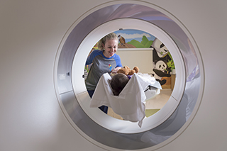 During a cardiac CT scan, an X-ray machine moves around your child's body in a circle to take a picture of each part of their heart. A computer then combines the pictures to make a three-dimensional (3D) picture of the whole heart.
Some CT scans might require that a dye is administered through your child's veins to provide contrast for the picture. Your child will need to lie very still during the test for at least 30 minutes, and younger patients sometimes need to be lightly sedated.
Learn why we use special CT scans for kids.
3D heart model printouts
For some patients, the Heart Institute at Children’s Colorado can use the information from a cardiac MRI, CT scan or cardiac catheterization along with three-dimensional (3D) printing technology to build a life-sized model of a patient’s heart. This allows us to understand your child’s heart in ways that were previously not possible.
This technique uses information collected during a CT scan, cardiac MRI or cardiac catheterization to build a virtual model, which is then used to build (or “print”) an exact physical model of your child’s heart. This is only available to patients who have a medical need for these tests.
Benefits of 3D printing the heart
When appropriate, we provide families with a model of your child’s heart to help you understand what the heart or blood vessels look like and what interventions are needed. Printing an exact anatomical model of a patient’s heart also allows us to:
- Understand abnormalities in the heart and blood vessels
- Provide counseling and education to eligible families before surgery
- Plan complex surgeries
- Plan interventions in the cardiac catheterization laboratory
- Educate medical trainees


Learn more about research and innovation at the Heart Institute.
Find locations with MRI and CT services.
Learn about other common heart tests.