- Doctors & Departments
-
Conditions & Advice
- Overview
- Conditions and Symptoms
- ¿Está enfermo su hijo?
- Parent Resources
- The Connection Journey
- Calma Un Bebé Que Llora
- Sports Articles
- Dosage Tables
- Baby Guide
-
Your Visit
- Overview
- Prepare for Your Visit
- Your Overnight Stay
- Send a Cheer Card
- Family and Patient Resources
- Patient Cost Estimate
- Insurance and Financial Resources
- Online Bill Pay
- Medical Records
- Política y procedimientos en el hospital
- Preguntamos Porque Nos Importa
-
Community
- Overview
- Addressing the Youth Mental Health Crisis
- Calendar of Events
- Child Health Advocacy
- Community Health
- Community Partners
- Corporate Relations
- Global Health
- Patient Advocacy
- Patient Stories
- Pediatric Affiliations
- Support Children’s Colorado
- Specialty Outreach Clinics
Your Support Matters
Upcoming Events
Colorado Hospitals Substance Exposed Newborn Quality Improvement Collaborative CHoSEN Conference (Hybrid)
lunes, 29 de abril de 2024The CHoSEN Collaborative is an effort to increase consistency in...
-
Research & Innovation
- Overview
- Pediatric Clinical Trials
- Q: Pediatric Health Advances
- Discoveries and Milestones
- Training and Internships
- Academic Affiliation
- Investigator Resources
- Funding Opportunities
- Center For Innovation
- Support Our Research
- Research Areas

It starts with a Q:
For the latest cutting-edge research, innovative collaborations and remarkable discoveries in child health, read stories from across all our areas of study in Q: Advances and Answers in Pediatric Health.


Cardiac Imaging with Echocardiogram (ECHO)
We are one of the largest programs in the country treating patients with heart problems from before birth through adulthood, with exceptional outcomes.


The Cardiac Imaging Program at Children's Hospital Colorado performs state-of-the-art heart tests for babies, kids and teens. Our team of heart imaging specialists can find out more about your child's heart by using tests like echocardiogram (ECHO), fetal echocardiogram (fetal ECHO), three-dimensional imaging (3D ECHO) and transesophageal echocardiogram (TEE).
Echocardiograms are performed in the Cardiology Clinic at our hospital on Anschutz Medical Campus in Aurora, South Campus in Highlands Ranch and at all satellite and Outreach Program locations.
We're also able to use telemedicine so that doctors outside of Denver (and even Colorado) can transmit these different types of echocardiographic studies to our team at Children's Colorado for review. This means that patients can now be examined closer to home and don't have to travel to our Denver Metro-area hospitals for these types of tests.
Echocardiogram (ECHO)
An echocardiogram (ECHO) is a non-invasive test that uses sound waves to create images — like an ultrasound of the heart. From this test, doctors can learn a lot about the structure and blood flow of the heart. An ECHO can show critical heart defects and structural or valve abnormalities in children of all ages. There is no known risk from exposure to ultrasound waves.
What to expect from an echocardiogram
- Your child will need to lie very still for 30 to 45 minutes; in some cases, very small children may need to be sedated by one of our dedicated cardiac anesthesiologists.
- A technician will put stickers on your child’s chest to monitor the heart rate, and your child will lie on their side or back for the test.
- The technician uses a wand with gel on it to get the images of your child’s heart (like the ultrasounds mothers get when pregnant). The room is dark and cozy, and your child is able to watch a movie while the test is being done.
- Sometimes a doctor will come in and review the test or ask for more photos. This is not necessarily a sign that anything is wrong – sometimes they just need more information.
Fetal echocardiogram (fetal ECHO)
A fetal echocardiogram is a safe ultrasound study performed on a pregnant mother's abdomen to show the structure of an unborn baby's heart. It provides valuable information on the growth and development of the baby's heart and blood vessels. Using this test, doctors specialized in fetal cardiology are able to diagnose a baby's structural heart problems and heart rhythm problems before birth. Learn more about our Fetal Cardiology Program for pregnant moms.
Three-dimensional echocardiogram (3D ECHO)
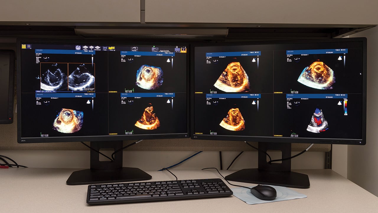
A three-dimensional echocardiogram, or 3D ECHO, lets doctors analyze heart function by producing a virtual 3D computer model of a child's heart, offering views of a heart’s anatomy that can’t be seen by standard echocardiogram. Because it helps cardiologist understand the structure and function of the heart in advance, it’s a crucial planning tool for surgery.
At Children’s Colorado, we pioneered 3D ECHO guidance for certain heart catheterization procedures, lowering and in some cases eliminating the need for patient exposure to X-ray radiation.
What to expect from a 3D ECHO
A 3D ECHO can be done at the same time as a standard ECHO, either at your child's bedside or in our outpatient clinic. It can be performed in children of all ages.
Transesophageal echocardiogram (TEE)
A transesophageal echocardiogram (TEE) is a special type of ECHO test that is used to assess the heart's function. It can be used during heart surgery or catheterization to help a surgeon see results of their operation in real time.
What to expect from a TEE
A TEE is a way to perform an echocardiogram by going through a patient's esophagus. A TEE is done either during heart surgery in the operating room or during a cardiac catheterization. Because the TEE is done while a patient is under anesthesia, your child will not feel the test or remember the procedure being done.
A TEE can be done in babies as small as four pounds. We also have a special three-dimensional TEE probe that allows detailed views of anatomy in older patients.
We are accredited by the Intersocietal Accreditation Commission
The Intersocietal Accreditation Commission (IAC) accredits imaging facilities and hospitals specific to echocardiography. IAC accreditation is a means by which facilities can evaluate and demonstrate the level of patient care they provide.


 720-777-0123


