Children's Hospital Colorado
Advances and Answers in Pediatric Health
U.S. News & World Report ranked in all 10 specialties badge

Advancing child health and pediatric specialty care through clinical discovery, multidisciplinary research and innovation

Improving the Evaluation of Fetal Arrhythmias with the InnV/Ao Doppler Technique

8/1/2018

A smiling blond woman in a blue shirt lies on an exam table. Dr. Howley looks at a screen on a computer-like machine showing fetal ultrasound results.

Key takeaways

  • The most widely used approaches for evaluating fetal heart rhythms have limits.

  • Our fetal cardiologists were the first to modify the superior vena cava and the ascending aorta (SVC/AAo) technique.

  • The new method, called the InnV/Ao Doppler technique, improves views of the retrograde atrial systolic flow and evaluation of fetal arrhythmia.


Research background: There are limitations in existing methods of assessing fetal arrhythmia

Fetal rhythm abnormalities, which include irregular fetal heart rates, occur in up to 2% of pregnancies and account for 10 to 20% of referrals to fetal cardiologists. Echocardiography is typically used to determine if the fetal heart arrhythmia is benign or if there is a pathological abnormality. During an echocardiogram, fetal heart rhythm is typically assessed using ultrasound tools called M-mode and pulsed spectral Doppler.

Simultaneously recording the pulsed Doppler flow profile in the superior vena cava (SVC) and the ascending aorta (AAo) is more accurate for atrioventricular (AV) and ventriculoatrial (VA) time measurements than M-mode.

Although this pulsed Doppler technique improves accuracy, there are several limiting factors. To be reliable in sampling the low velocity SVC atrial flow and simultaneous AAo, the Doppler signal needs an ideal angle of insonation (approaching 0 degrees) and low wall motion filter (cannot be set too high). Otherwise, the A-wave in the SVC may not be detected.

Research methods: Modifying the way we evaluate arrhythmia in fetus

The team in the Fetal Cardiology Program at the Colorado Fetal Care Center at Children's Hospital Colorado, modified the SVC/AAo technique to improve the examination of fetal arrhythmias and evaluating fetal arrhythmias. They simultaneously recorded the pulsed wave Doppler signals in the innominate vein (InnV) and transverse aortic arch (Ao) from an axial view of the fetal thorax.


By employing a four-chamber view in a horizontal position, the InnV is easily identified by sweeping cephalad to the three-vessel trachea view. The InnV is located immediately superior to the transverse arch, and can be positioned in parallel to the ultrasound beam for clear and precise spectral Doppler sampling of the venous A-wave signal.

Research results: An improved view of the retrograde atrial systolic flow

When the Doppler sample volume is widened, blood velocities in both the InnV and the Ao can be simultaneously recorded. (See Figure 1.)
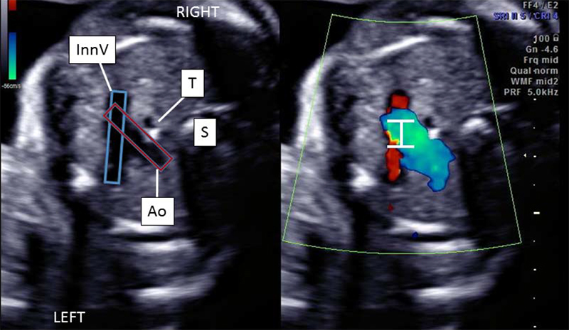
A black and white, axial 2D and color Doppler image of the fetal thorax demonstrating the position and relationship of the innominate vein (labeled InnV), the transverse aortic arch (labeled Ao), the trachea (labeled T) and the spine (labeled S).
Figure 1: a) Axial 2D and color Doppler image of the fetal thorax demonstrating the position and relationship of the innominate vein (InnV), the transverse aortic arch (Ao), the trachea (T) and the spine (S). The sample gate is placed simultaneously over the InnV and the Ao to determine the mechanical relationship between the atria and the ventricles respectively. 
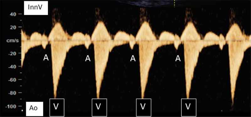
A graph-like image of a fetus in sinus rhythm with a normal AV interval of 125 ms. There is a y-axis of cm/s labeled from -100 to 40. There is a yellowish wave along x-axis.
Figure 1: b) A fetus in sinus rhythm with a normal AV interval of 125 ms.

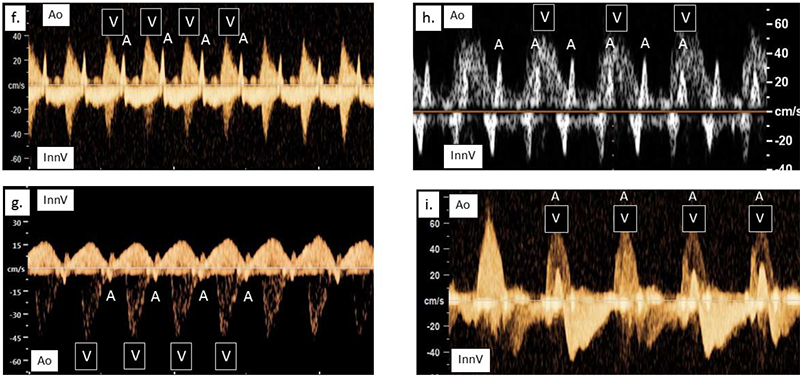
The InnV/Ao method provides a near 0-degree angle of insonation of the InnV to optimize visualization of the retrograde atrial systolic flow. (See Figure 2 for fetal arrhythmias detected with the method.)
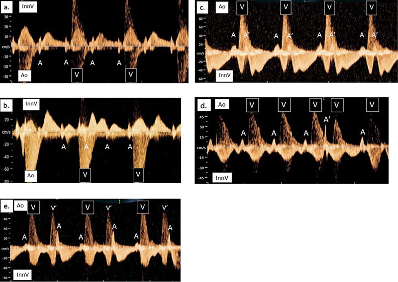
Five simultaneous recordings of the InnV and the Ao in different fetal arrhythmias. There are 5 graph-like images where there is a y-axis of cm/s labeled from -100 to 40 and yellowish waves along x-axis.
Figure 2: Simultaneous recordings of the InnV and the Ao in different fetal arrhythmias. (A, atrial systole; A’, premature atrial contraction; V, ventricular systole; V’, premature ventricular contraction; InnV, innominate vein; Ao, transverse aortic arch; R-R interval, cardiac cycle length; ms, milliseconds). Bradycardia a). A fetus in type 2, 2° AV block. The atrial rate is regular (A-A interval = 400 ms). Every other atrial beat is conducted to the ventricle; the R-R interval is 800 ms. b). Autoantibody-mediated complete heart block with absence of fetal AV synchrony. The atrial rate is regular (A-A interval = 414 ms) and the R-R interval is 976 ms. c). A fetus with blocked atrial bigeminy. The atrial rate is regularly irregular with a long- short-long pattern (A-A’ interval = 233 ms, A’-A interval 486 ms). The ventricular rate is regular with an R-R interval of 697 ms. Irregular Rhythm d). Isolated conducted premature atrial contraction (A’). e). Ventricular bigeminy with a premature ventricular contraction (V’) every other beat. In contrast to premature atrial beats, the ventricular signal occurs prior to the atrial signal (A).
Four graph-like images where there is a y-axis of cm/s labeled from -100 to 40 and yellowish waves along x-axis each portraying different fetal heart rhythms.
Figure 2: Tachycardia f). 1:1 reciprocating supraventricular tachycardia (SVT) with a short VA interval, (VA= 103 ms and AV = 160 ms). The ventricular rate is regular with an R-R interval of 260 ms. g). 1:1 reciprocating SVT with a long VA interval, (VA= 190 ms and AV = 130 ms). The ventricular rate is regular with an R-R interval of 321 ms. h). Atrial flutter with 2:1 AV conduction. The atrial rate is regular (A-A interval = 128 ms) and the R-R interval is 256 ms. i). Junctional ectopic tachycardia with simultaneous depolarization of the atria (A) and ventricles (V).

Research conclusion: Our InnV/Ao Doppler technique is an acceptable modification of the SVC/AAo technique

Children's Colorado is the first to use simultaneous Doppler recordings of the InnV and Ao from an axial plane to assess fetal cardiac arrhythmias. The InnV/Ao Doppler technique is an accurate method to assess fetal heart rhythm and can be regularly performed in the clinical setting.