720-777-0123


Key takeaways
-
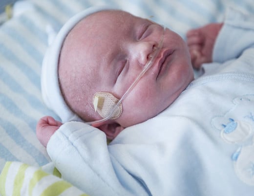
Newborn screening for congenital heart disease is not always performed by a pediatrics-trained sonographer, especially in smaller community hospitals.
-
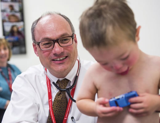
Pediatric echocardiography experts at Children’s Colorado helped create recommendations for adult sonographers to detect C-CHD in newborns to improve outcomes.
-
The recommendations should help accurately identify and quickly stabilize more newborns with C-CHD, reducing morbidity and mortality.
Background for recommendations: importance of early detection of critical congenital heart disease
Congenital heart defects (CHD) are the leading cause of death and severe illness in infants in developed countries. For newborns diagnosed with the most serious type of CHD, C-CHD, surgery or catheter intervention is necessary before an infant is one year old. Despite it being one of the most common types of congenital malformations, C-CHD is not always diagnosed early and referred to a pediatric cardiologist. Factors like the type of C-CHD lesion and geographic location affect detection rates, and not all hospitals have pediatric-trained sonographers.
Early detection can save lives
With some symptoms not appearing until after the first 48 hours of life, early detection of C-CHD is critical for staging the appropriate intervention. A delayed or missed diagnosis can lead to death or severe and long-lasting health problems such as severe cyanosis, cardiovascular collapse, or neurodevelopmental abnormalities due to brain injury.
There is a critical window of time during newborn hospitalization where screening for and detection of C-CHD could lead to improved outcomes. Since adult sonographers typically don’t screen for C-CHD, specific guidelines and standards could aid in their detection of C-CHD.
Members of the American Society of Echocardiography, including experts in the Heart Institute at Children’s Hospital Colorado, created recommendations for adult sonographers that describe the pathology, along with 30 videos demonstrating the 12 most common forms of C-CHD that are detectable by Pulse Oximetry Screening (POS).
C-CHD screening methodology and pulse oximetry screening
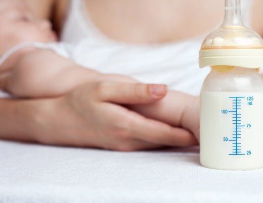
Common current C-CHD screening methodology focuses on POS, a non-invasive way to detect hypoxemia. Previous research from the American Heart Association and the American Academy of Pediatrics found that when used within the first 24 hours of life, POS can detect C-CHD, the estimated sensitivity for detecting C-CHD was 69.6%, while specificity was 99% and the positive predictive value was 47%.
Many types of C-CHD are targeted for their reliability of identification by POS.

A failed POS test mandates immediate evaluation for C-CHD, with echocardiography the diagnostic modality of choice. Access to pediatric echocardiography and pediatric cardiology services may be limited in rural areas or small hospitals, and the accuracy of interpretation by adult cardiologists is significantly lower.
Recommended infrastructure for adult cardiac sonographers
When screening for C-CHD in newborns, adult cardiac sonographers should ensure the following infrastructure are present:
- Age-appropriate echo equipment
- A mechanism for storage and transmission of images
- A structured, open and fast communication process between referring provider, sonographer and reading physician that begins in the newborn nursery or neonatal intensive care unit (NICU)

Instrumentation and patient setting
Newborn echocardiograms are best performed with a variety of probes with a range of frequencies.
The video screen and display should be of suitable size and quality for observation and interpretation and identify or include:
- Performing institution
- Appropriate patient identifiers
- Date and time of the study
- Range or depth markers
- Measurement capabilities
- Distance between two points
- Area on the 2D image
- Blood flow velocities
- Time intervals
- Peak and mean gradients from spectral Doppler studies
- Optimized frame rate for adequate visualization of anatomy at higher neonatal heart rates
Communication with pediatric cardiologist or referral center
Centers performing screening echocardiograms in newborns should have a formal relationship with a physician or referral center with expertise in C-CHD and develop a smooth process for communication:
-
- As soon as decision is made to obtain the test, the performing site's newborn nursery or neonatal intensive care unit notifies receiving site of a pending echocardiogram to review.
- Receiving sites may provide a form to performing sites to accompany the echocardiogram transmission, including:
- Demographic information
- Indication for the study
- Patient height and weight
- Concurrent systemic blood pressure
- Desired urgency of interpretation
- Contact information so results can be called back to the referring provider
- Referring provider should communicate directly with the reading physician if there is a particular sense of urgency or patient acuity.
- After review by reading physician, results go to performing site securely and efficiently.
- A formal policy for communicating results, starting with a phone call to referring provider to relay results and allow for discussion.
- The interpreting pediatric cardiologist should work with the referring center to develop a method to relay a final report.
- Finally, open lines of communication should exist between echocardiography labs at both hospitals.
Specific imaging recommendations
Imaging recommendations include:
- Pediatric-specific imaging protocols such as taking longer (10 to 20 second) videos
- Complete views of the heart that show any base or apex abnormalities and relational orientation of the cardiac anatomy
- The sonographer should be familiar with typical signs of C-CHD that show up on echos and promptly alert the appropriate physician.
Conclusion: increased C-CHD detection and accuracy improves outcomes and saves lives
Now that POS is mandatory in newborns prior to discharge from the nursery, more community hospitals are performing echocardiograms on newborns to screen for C-CHD. Partnerships between hospitals performing newborn delivery and care and offsite pediatric cardiology experts available to interpret newborn screening echocardiography studies and perform exams when needed are critical to ensure the best outcomes.
These recommendations, when used, should allow more newborns with C-CHD to be accurately identified and stabilized quickly, reducing the morbidity and mortality in this at-risk population. They will also help sonographers not fully trained in pediatric echocardiography to obtain images that allow for accurate diagnosis (or exclusion) of C-CHD.